
Imagine walking into a doctor's office, putting on a lightweight headset, and instantly seeing a living, breathing, three-dimensional holographic map of the organs inside a patient’s body. It sounds like pure science fiction—something pulled straight from a Hollywood superhero movie.
But on June 10, 2026, researchers at the Massachusetts Institute of Technology (MIT) turned this exact concept into a reality.
Published in Nature Communications Engineering, a team of MIT engineers unveiled a major medical breakthrough: AR-VIU (Augmented Real-Time Volumetric Imaging in Ultrasound). This new technology completely reimagines how we look inside the human body, transforming a notoriously difficult medical skill into something almost anyone can understand.
The Hidden Challenge of Traditional Ultrasounds
Ultrasound technology is one of the safest and most vital diagnostic tools we have. It uses harmless sound waves to peer inside the body without exposing patients to radiation. However, it has a massive, hidden flaw: it is incredibly hard to read.
When a technician glides a traditional ultrasound wand over your skin, the machine spits out flat, grainy, two-dimensional cross-sectional slices on a monitor. The technician then has to use intense mental gymnastics to piece those 2D puzzle pieces together into a 3D picture inside their head.
Because of this steep learning curve, mastering an ultrasound can take years of rigorous training. If you don't have that experience, looking at an ultrasound monitor just feels like staring at moving static.
Enter AR-VIU: The Power of Holographic Medicine
The MIT team, led by graduate students Jason Hou and Shrihari Viswanath alongside senior author Canan Dagdeviren, set out to eliminate that mental burden entirely.
Instead of forcing the human brain to reconstruct a 3D image, they built a system that does it automatically. By combining an innovative hardware design with cutting-edge augmented reality (AR), the AR-VIU system gives medical operators literal "X-ray vision."
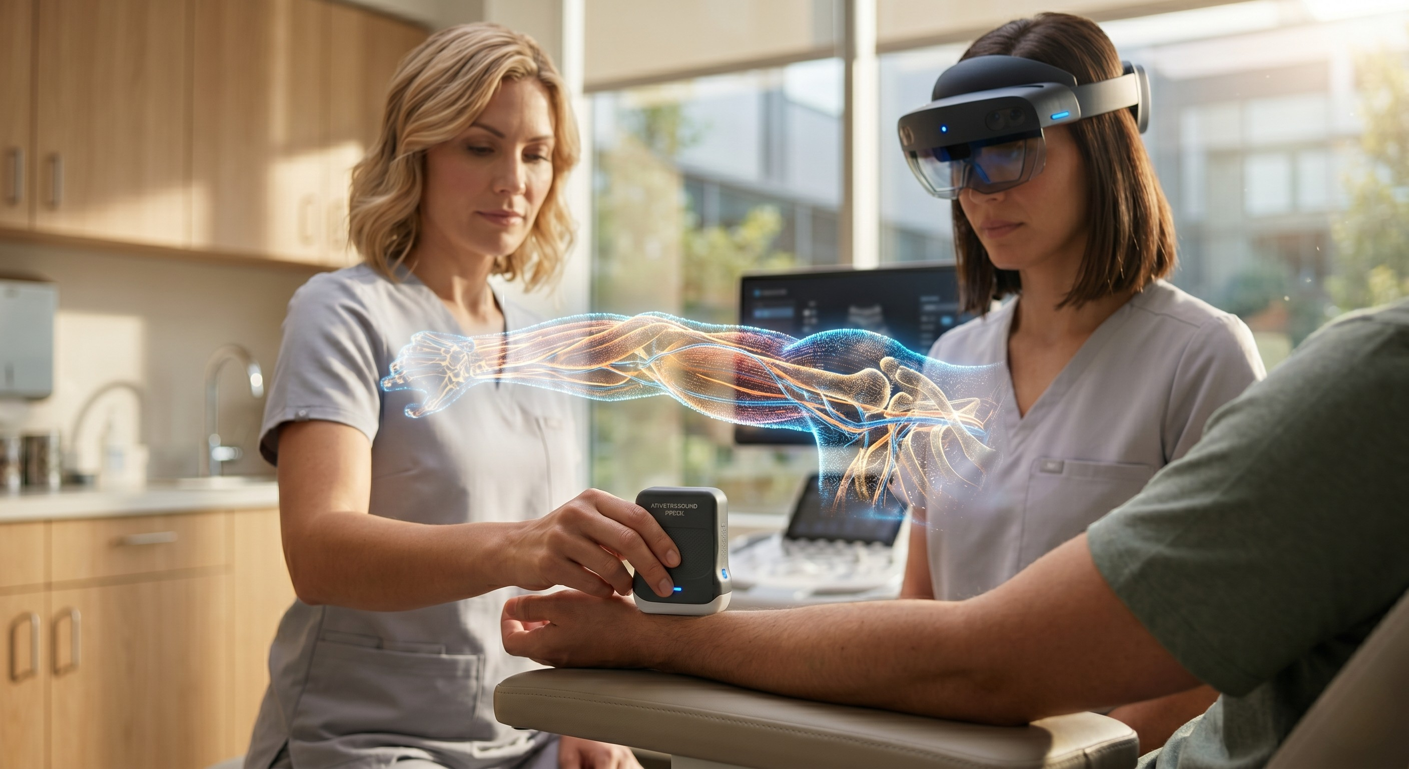
When wearing an AR headset, the medical professional no longer looks away at a flat screen. Instead, a precise, real-time 3D digital model of the internal tissue is projected directly onto the patient’s body, perfectly aligned with their anatomy.
How Traditional Scans Compare to MIT's Innovation
| Feature | Traditional 2D Ultrasound | MIT's AR-VIU System |
| Display Type | Flat, abstract 2D slices on a monitor | Real-time, 3D holographic overlay |
| Cognitive Effort | High; requires mental 3D assembly | Low; highly intuitive spatial context |
| Learning Curve | Long, steep curve for specialists | Rapid; beginners perform like veterans |
| Hardware Profile | Bulky, high-power, expensive machinery | Pocket-sized probe, low-power architecture |
How Does It Work? (Gaming Tech Meets Healthcare)
The brilliance of the AR-VIU system relies on two main components working together seamlessly:
- The Pocket-Sized Probe: The hardware is a compact ultrasound probe slightly smaller than a deck of cards. Inside, the ultrasound sensors are arranged in an innovative, empty square shape. This "ultra-sparse" design allows the device to capture rich 3D volumetric data using far fewer components, making it vastly cheaper to build and less power-hungry than old-school machinery.
- The Unreal Engine Software: Raw data captured by the probe is instantly compressed and streamed into Unreal Engine—the exact same high-powered 3D computer graphics software used to build hyper-realistic video games like Fortnite. Unreal Engine translates the raw data into a live 3D structure that an operator can view from different angles simply by tilting their head.
The Impact: Turning Novices into Experts
To see if this system actually worked in practice, MIT conducted a controlled study. They tasked both seasoned experts and complete novices with finding and mapping deep objects hidden inside artificial tissue blocks (simulating tasks like locating a tumor for a needle biopsy).
The results were staggering. When using a traditional 2D monitor, the experts completely outpaced the beginners. But when the beginners put on the AR headset and used the AR-VIU system, the performance gap virtually vanished.
First-time users were able to track and identify deep-tissue features almost as accurately and quickly as veteran sonographers.
"The 3D system imposes less brain drain, it's more intuitive, and it's easier to understand what is happening in the targeted region," noted senior author Canan Dagdeviren.
Democratizing Care: Why This Matters for Everyone
The true long-term magic of this breakthrough isn't just that it looks cool; it is the democratization of medicine.
Because the hardware is inexpensive and the software makes it incredibly easy to use, this tech can easily leave the major university hospitals and head out into the real world.
Imagine emergency EMTs using this in the back of a moving ambulance to check for internal bleeding, or rural clinic nurses using it to accurately place IV needles or perform biopsies without needing an on-call imaging specialist. By taking the "brain drain" out of ultrasound interpretation, MIT has paved the way for faster, safer, and more accessible healthcare for everyone.
.png?width=160&height=90&name=cdn%20(1).png "cdn (1)")